. 2021;80(2):761-774.
doi: 10.3233/JAD-200919.

Particulate Matter Exposure Exacerbates Amyloid-β Plaque Deposition and Gliosis in APP/PS1 Mice

Affiliations

Particulate Matter Exposure Exacerbates Amyloid-β Plaque Deposition and Gliosis in APP/PS1 Mice

Bijayani Sahu et al. J Alzheimers Dis. 2021.

Abstract

Background: Alzheimer's disease (AD) is a progressive neurodegenerative disorder characterized by the accumulation of amyloid-β (Aβ) plaques, neuroinflammation, and neuronal death. There are several well-established genetic and environmental factors hypothesized to contribute to AD progression including air pollution. However, the molecular mechanisms by which air pollution exacerbates AD are unclear.

Objective: This study explored the effects of particulate matter exposure on AD-related brain changes using the APP/PS1 transgenic model of disease.

Methods: Male C57BL/6;C3H wild type and APP/PS1 mice were exposed to either filtered air (FA) or particulate matter sized under 2.5μm (PM2.5) for 6 h/day, 5 days/week for 3 months and brains were collected. Immunohistochemistry for Aβ, GFAP, Iba1, and CD68 and western blot analysis for PS1, BACE, APP, GFAP, and Iba1 were performed. Aβ ELISAs and cytokine arrays were performed on frozen hippocampal and cortical lysates, respectively.

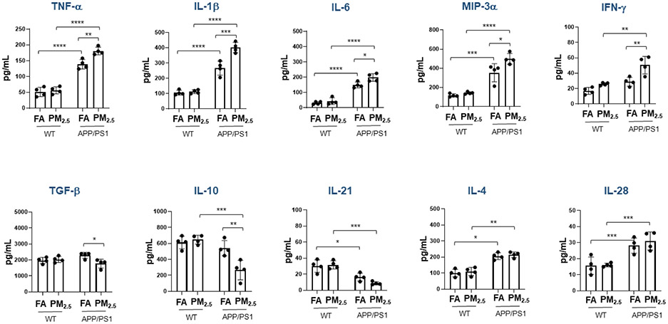
Results: The Aβ plaque load was significantly increased in the hippocampus of PM2.5-exposed APP/PS1 mice compared to their respective FA controls. Additionally, in the PM2.5-exposed APP/PS1 group, increased astrocytosis and microgliosis were observed as indicated by elevated GFAP, Iba1, and CD68 immunoreactivities. PM2.5 exposure also led to an elevation in the levels of PS1 and BACE in APP/PS1 mice. The cytokines TNF-α, IL-6, IL-1β, IFN-γ, and MIP-3α were also elevated in the cortices of PM2.5-exposed APP/PS1 mice compared to FA controls.

Conclusion: Our data suggest that chronic particulate matter exposure exacerbates AD by increasing Aβ plaque load, gliosis, and the brain inflammatory status.

Keywords: Alzheimer’s disease; amyloid-β plaques; neuroinflammation; particulate matter.


Conflict of interest statement

Disclosure

The authors have no actual or potential conflicts of interest.

Figures
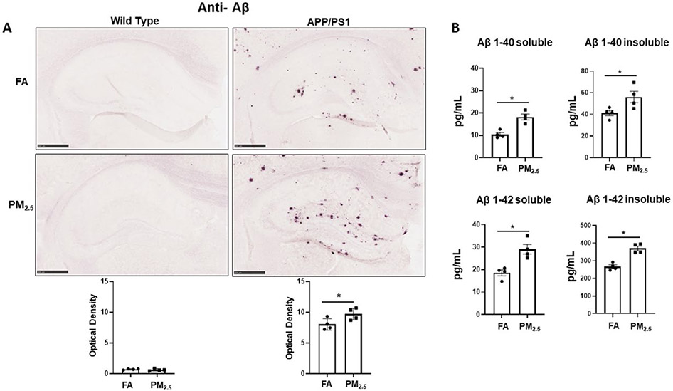
Fig. 1.
Fig. 1.. Particulate matter inhalation increased Aβ plaque load in APP/PS1 hippocampus.
Male littermate control C57BL/6;C3H wild type and APP/PS1 mice were exposed to FA or PM2.5 for 3 months and brains were collected, sectioned and immunostained with anti-Aβ antibody using Vector VIP as the chromogen. (A) Representative immunohistochemical staining images (5x) of the hippocampus for Aβ are shown. Quantification of immunoreactivity was performed, and optical densities obtained were averaged and graphed as mean ± SEM (n=4), *p < 0.05, scale bar 500 μm. (B) The levels of both soluble and insoluble amyloid-beta 1-40 (Aβ 1-40) and 1-42 (Aβ 1-42), were measured in hippocampi from wild type and APP/PS1 mice using ELISAs. Results are shown as mean ± SEM (n=4), *p < 0.05.
Fig. 2.
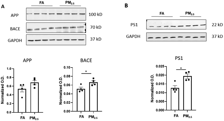
Fig. 2.. Particulate matter inhalation increased APP processing enzyme levels in APP/PS1 hippocampus.
Frozen hippocampi from the APP/PS1 mice exposed to FA or PM2.5 were lysed and resolved via SDS-PAGE and western blots performed using (A) anti-APP, anti-BACE, and (B) anti-PS1 antibodies. Anti-GAPDH antibodies were used to probe the respective blots as a loading control. Quantification shows arbitrary densitometry units of each protein signal normalized to its respective GAPDH loading control. Results are shown as mean ± SEM (n=4), *p < 0.05.
Fig. 3.
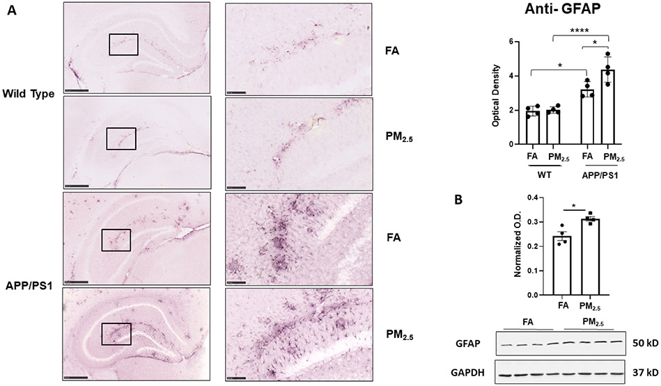
Fig. 3.. Particulate matter inhalation increased astrogliosis in APP/PS1 hippocampus.
Male littermate control C57BL/6;C3H wild type and APP/PS1 mice were exposed to FA or PM2.5 for 3 months and brains were collected, sectioned and immunostained with anti-GFAP antibody using Vector VIP as the chromogen. Representative immunohistochemical staining images (5x and 20x) of the hippocampus are shown (A). Quantification of immunoreactivity was performed, and optical densities obtained were averaged and graphed as mean ± SEM (n=4), *p < 0.05 and ****p < 0.0001; scale bar 500 μm. (B) Frozen hippocampi from the mice were lysed and resolved via SDS-PAGE and western blots performed using anti-GFAP and anti-GAPDH antibodies. Quantification shows arbitrary densitometry units of protein signal normalized to its respective GAPDH loading control. Results are shown as mean ± SEM (n=4), *p < 0.05.
Fig. 4.
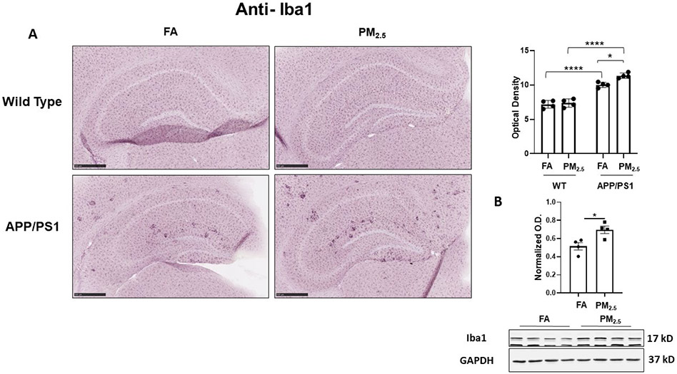
Fig. 4.. Particulate matter inhalation increased microgliosis in APP/PS1 hippocampus.
Male littermate control C57BL/6;C3H wild type and APP/PS1 mice were exposed to FA or PM2.5 for 3 months and brains were collected, sectioned and immunostained with anti-Iba1 antibody using Vector VIP as the chromogen. Representative immunohistochemical staining images (5x) of the hippocampus are shown (A). Quantification of immunoreactivity was performed and optical densities obtained were averaged and graphed as mean ± SEM (n=4), *p < 0.05 and ****p < 0.0001; scale bar 500 μm. (B) Frozen hippocampi from the mice were lysed and resolved via SDS-PAGE and western blots performed using anti-Iba1 and anti-GAPDH antibodies. Quantification shows arbitrary densitometry units of protein signal normalized to its respective GAPDH loading control. Results are shown as mean ± SEM (n=4), *p < 0.05.
Fig. 5.
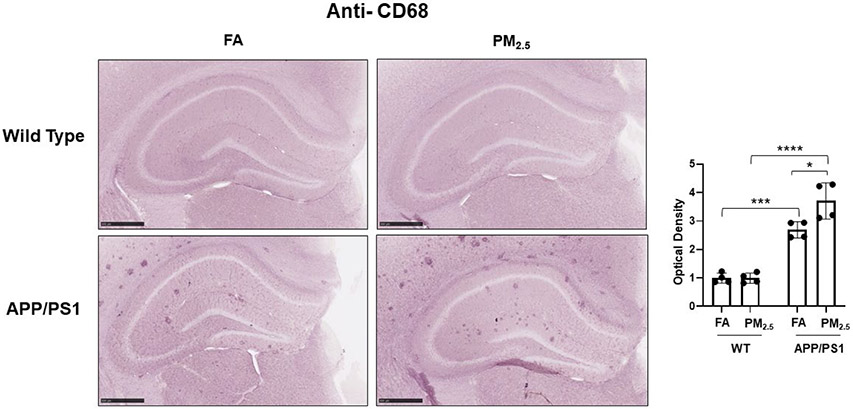
Fig. 5.. Particulate matter inhalation increased microglial phagocytic phenotype in APP/PS1 hippocampus.
Male littermate control C57BL/6;C3H wild type and APP/PS1 mice were exposed to FA or PM2.5 for 3 months and brains were collected, sectioned and immunostained with anti-CD68 antibody using Vector VIP as the chromogen. Representative immunohistochemical staining images (5x) are shown. Quantification of immunoreactivity was performed and optical densities obtained were averaged and graphed as mean ± SEM (n=4), *p < 0.05, ***p < 0.001 and ****p < 0.0001; scale bar 500 μm.
Fig. 6.
Fig. 6.. Particulate matter inhalation minimally affected brain cytokines in wild type mice.
Male littermate control C57BL/6;C3H wild type and APP/PS1 mice were exposed to FA or PM2.5 for 3 months and brains were collected. Temporal cortices of wild type and APP/PS1 mice were lysed and used for a slide-based mouse cytokine array. Results from wild type and APP/PS1 brains are shown as mean ± SEM (n=4), *p < 0.05, **p < 0.01, ***p < 0.001 and ****p < 0.0001.

References

    1. Bondi MW, Edmonds EC, Salmon DP (2017) Alzheimer's Disease: Past, Present, and Future. J Int Neuropsychol Soc 23, 818–831. - PMC - PubMed
    1. Hardy J, Selkoe DJ (2002) The amyloid hypothesis of Alzheimer's disease: progress and problems on the road to therapeutics. Science 297, 353–356. - PubMed
    1. Jia JP, Meng R, Sun YX, Sun WJ, Ji XM, Jia LF (2005) Cerebrospinal fluid tau, Abeta1-42 and inflammatory cytokines in patients with Alzheimer's disease and vascular dementia. Neurosci Lett 383, 12–16. - PubMed
    1. Querfurth HW, LaFerla FM (2010) Alzheimer's disease. N Engl J Med 362, 329–344. - PubMed
    1. Welge V, Fiege O, Lewczuk P, Mollenhauer B, Esselmann H, Klafki HW, Wolf S, Trenkwalder C, Otto M, Kornhuber J, Wiltfang J, Bibl M (2009) Combined CSF tau, p-tau181 and amyloid-beta 38/40/42 for diagnosing Alzheimer's disease. J Neural Transm (Vienna) 116, 203–212. - PubMed

Publication types

MeSH terms

LinkOut - more resources