. 2016 Sep 27;113(39):10797-801.
doi: 10.1073/pnas.1605941113. Epub 2016 Sep 6.

Magnetite pollution nanoparticles in the human brain

Affiliations

Magnetite pollution nanoparticles in the human brain

Barbara A Maher et al. Proc Natl Acad Sci U S A. .

Abstract

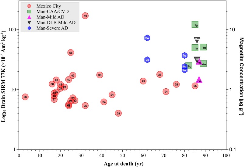
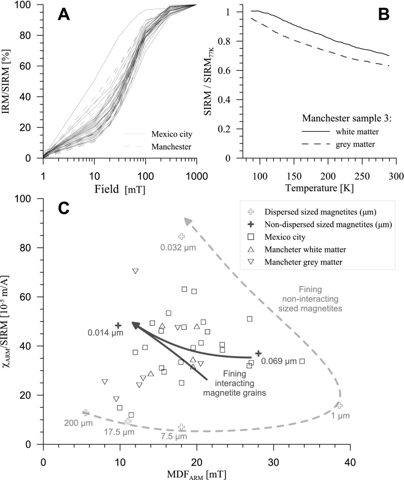
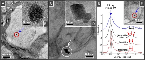
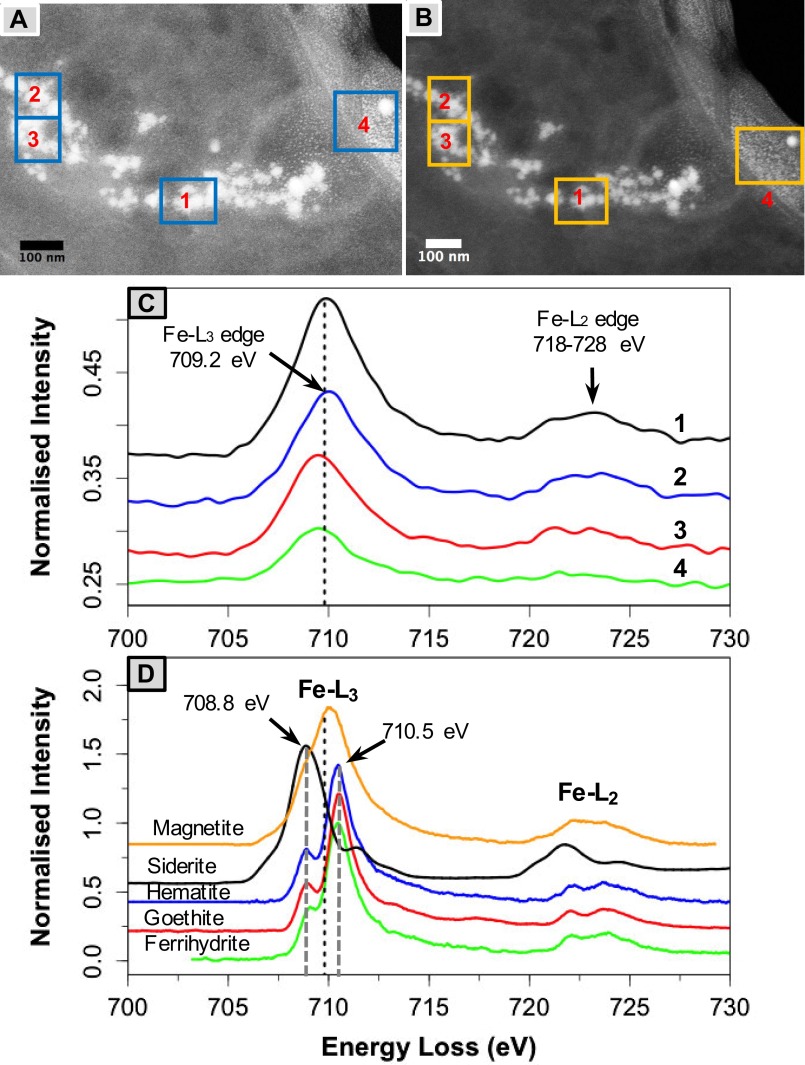
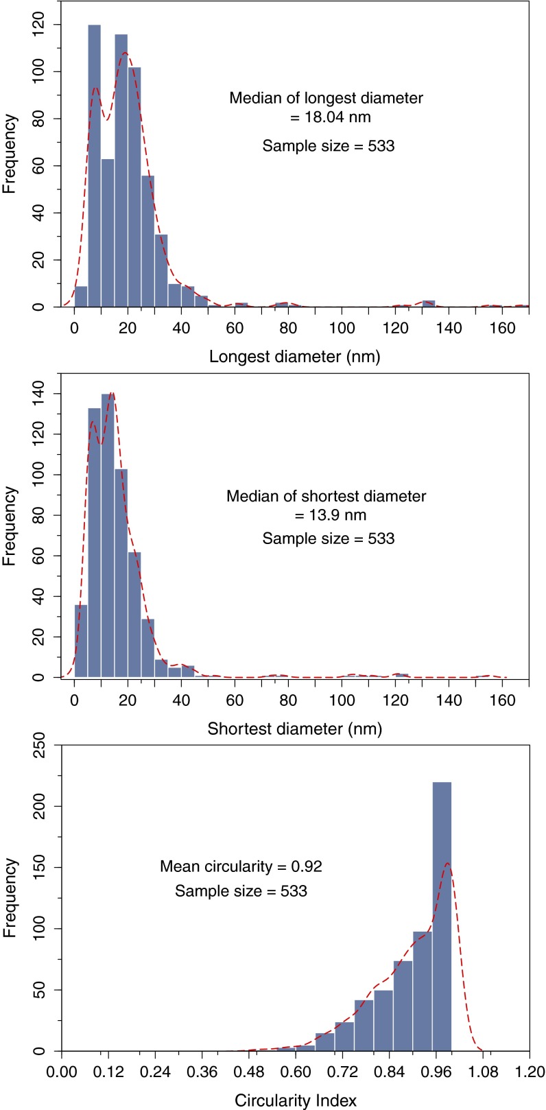
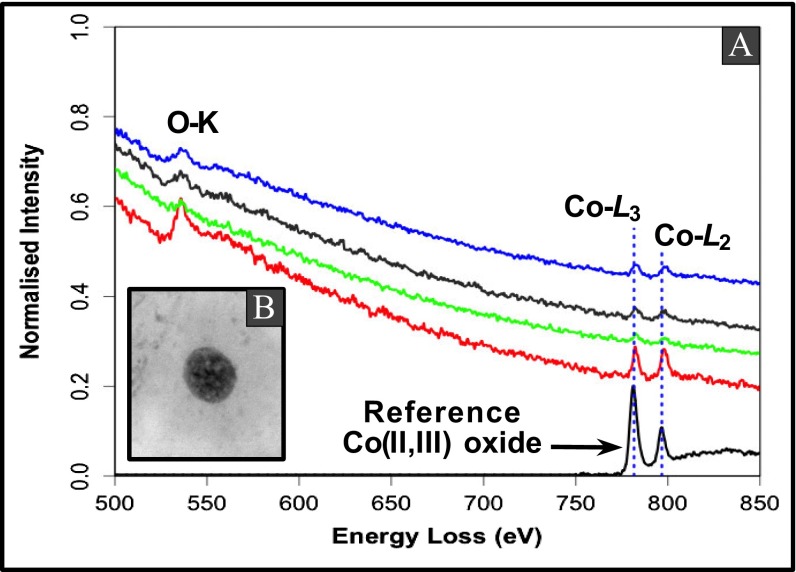
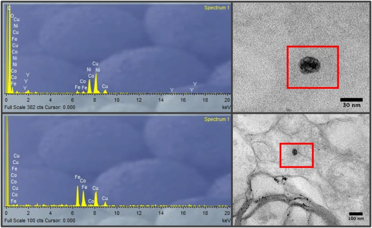
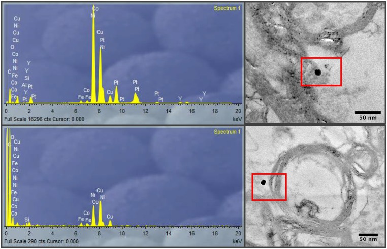
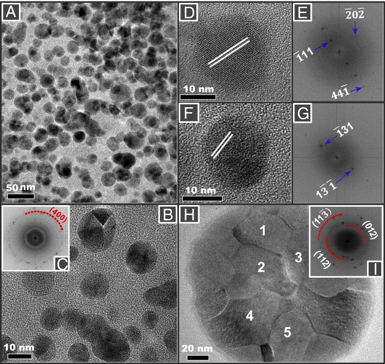
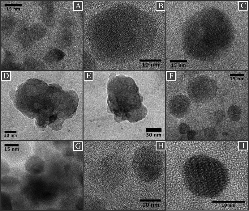
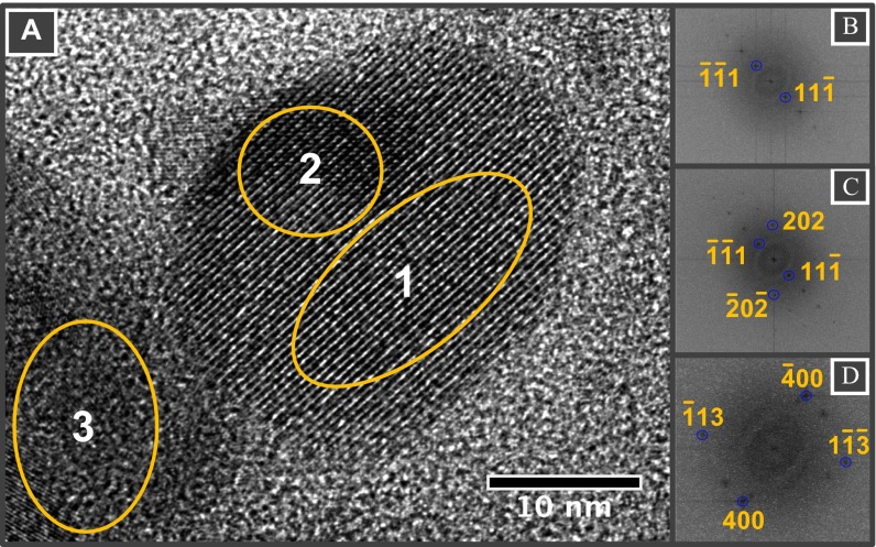
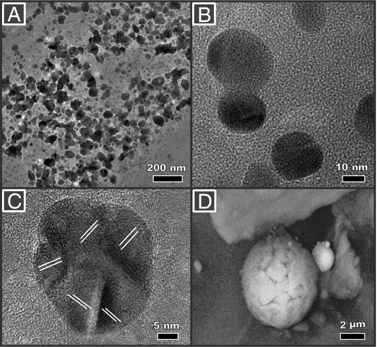
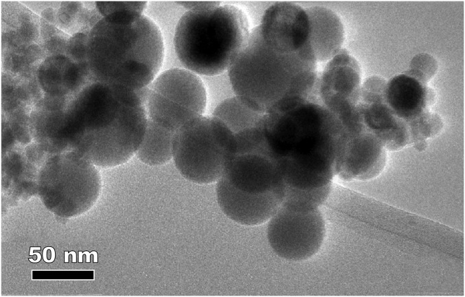
Biologically formed nanoparticles of the strongly magnetic mineral, magnetite, were first detected in the human brain over 20 y ago [Kirschvink JL, Kobayashi-Kirschvink A, Woodford BJ (1992) Proc Natl Acad Sci USA 89(16):7683-7687]. Magnetite can have potentially large impacts on the brain due to its unique combination of redox activity, surface charge, and strongly magnetic behavior. We used magnetic analyses and electron microscopy to identify the abundant presence in the brain of magnetite nanoparticles that are consistent with high-temperature formation, suggesting, therefore, an external, not internal, source. Comprising a separate nanoparticle population from the euhedral particles ascribed to endogenous sources, these brain magnetites are often found with other transition metal nanoparticles, and they display rounded crystal morphologies and fused surface textures, reflecting crystallization upon cooling from an initially heated, iron-bearing source material. Such high-temperature magnetite nanospheres are ubiquitous and abundant in airborne particulate matter pollution. They arise as combustion-derived, iron-rich particles, often associated with other transition metal particles, which condense and/or oxidize upon airborne release. Those magnetite pollutant particles which are <∼200 nm in diameter can enter the brain directly via the olfactory bulb. Their presence proves that externally sourced iron-bearing nanoparticles, rather than their soluble compounds, can be transported directly into the brain, where they may pose hazard to human health.

Keywords: Alzheimer's disease; airborne particulate matter; brain magnetite; combustion-derived nanoparticles; magnetite pollution particles.


Conflict of interest statement

The authors declare no conflict of interest.

Figures

Fig. S1.
Fig. S1.
SIRM 77 K (10−6 A m2/kg) and estimated magnetite concentration (micrograms per gram) for frontal cortex samples versus age at death, Mexico City and Manchester cases. The annual mean airborne PM2.5 concentration (micrograms per cubic meter) is given for the residence area of the Mexican cases (inside each data symbol); SIRM values for gray (g) and white (w) matter are given for the Manchester cases, together with their clinical diagnosis upon death (CAA, cerebral amyloid angiopathy; CVD, cerebrovascular disease; DLB, dementia with Lewy bodies; see Tables S1 and S2).
Fig. S2.
Fig. S2.
Magnetic analyses of brain tissue samples (freeze-dried): (A) acquisition of isothermal (RT) remanent magnetization in applied DC fields from 5 mT to 1 T. All samples acquire most of their magnetization at fields < 100 mT, indicating the dominant presence of ferrimagnetic minerals (e.g., magnetite and/or maghemite). The magnetically softest sample (the Mexico City case to the left of all remaining samples) has the highest SIRM value (case 282). (B) Measurement of LT remanence (77 K, DC field 1 T) upon warming to RT, showing the thermal unblocking of the superparamagnetic particles. (C) Comparison between the brain samples and sized, synthetic magnetites of known grain size and degree of dispersion (37), as measured by the RT ARM, normalized by the SIRM, plotted against the median destructive field of the ARM (MDFARM, in milliteslas). All of the measurable brain samples fall within the region of the least-dispersed synthetic, submicrometer magnetites, indicating magnetic interactions, and hence agglomeration/clustering of some of the brain magnetite particles.
Fig. 1.
Fig. 1.
Transmission electron micrographs of brain thin sections, identifying two distinct types of magnetite morphologies within frontal cells: (A and F) rounded particles (A shown at higher magnification in B); and (C) angular, euhedral particles, which we attribute to endogenous formation (particles from C shown at higher magnification in D). (E) EELS spectra (in blue) for the rounded particle shown in F and for standard iron oxide species. The position of the Fe−L3 edge absorption peak, the broad feature of the Fe−L2 (compared with the sharp edges, arrowed, of the fully oxidized Fe3+ phases), and the integrated areas of the L3/L2 (5.5) and the Fe/O (0.56) are all consistent with magnetite (also see Figs. S3 and S4).
Fig. S3.
Fig. S3.
(A) High-angle annular diffraction and (B) dark-field TEM micrographs showing spherical magnetic nanoparticles in brain tissues. (C) Fe−L2,3 EELS spectra of nanoparticles identified in the selected areas (boxes 1 through 4) showing the absence of any preedges (see hematite, goethite, and ferrihydrite preedge at ∼708.8 eV), Fe−L3 edges centered at 708 eV, and broad Fe−L2 features characteristic of magnetite, compared with the Fe−L2,3 EELS spectra (D) of standard magnetite, siderite, hematite, goethite, and two-line ferrihydrite.
Fig. S4.
Fig. S4.
Fe−L2,3-edge spectra of magnetic particles found in brain samples. The Fe−L3 and Fe−L2 edges in all three samples are at 708.7 to 709.8 eV and 72 to 723 eV, in excellent agreement with the chemical shift in EELS spectra for the magnetite structure (also see Fig. S3).
Fig. S5.
Fig. S5.
Particle size distribution of magnetic particles in brain magnetic extracts. Particle size measurements were carried out on all of the HRTEM micrographs collected from six brain magnetic extracts from different subjects. The ImageJ software package was used to describe the imaged particles (spherical and nonspherical) in terms of the longest and shortest diameters, perimeter projected area, or equivalent spherical diameter.
Fig. S6.
Fig. S6.
(A) Co−L2,3 EELS spectra of cobalt (II, III) oxide nanoparticles associated with magnetite particles in brain tissues. Co−L3 and Co−L2 edges from different areas of a brain tissue sample (B) are centered at ∼780 and ∼796 eV, respectively, in a good match with an EELS spectrum of a standard cobalt (II,III) oxide.
Fig. S7.
Fig. S7.
EDX analysis of metal-bearing NPs in brain tissue samples, showing presence of Fe, Ni, and Co (and possibly Cu, with the caveat that the samples were mounted on holey carbon films on Cu grids).
Fig. S8.
Fig. S8.
EDX analysis of metal-bearing NPs in brain tissue samples, showing the presence of Fe, Ni, Pt, Co, and, possibly, Cu.
Fig. 2.
Fig. 2.
Transmission electron micrographs of rounded particles magnetically extracted from human brain samples: (A, D, F, and H) Mexico City cases; (B) Manchester case. (H) A large (∼150-nm diameter) spherical particle with fused, interlocking magnetite/maghemite surface crystallites. (C, E, and G) Indexing of the lattice fringes of the brain particles is consistent with the (400) reflection of magnetite and (I) mixed magnetite and maghemite of selected areas 1–5 in H.
Fig. S9.
Fig. S9.
(AI) A collection of HRTEM micrographs of magnetite particles, extracted from brain tissues, showing dominant rounded morphologies. (C) Micrograph shows fused magnetite particles, and (D and E) micrographs show aggregated magnetite particles.
Fig. S10.
Fig. S10.
(A) HRTEM micrograph of magnetically extracted magnetite particles from brain tissues. (BD) FFT patterns of selected areas (1, 2, and 3, respectively) featuring a single crystal (B) and magnetite particles superimposed at ∼90° (C and D).
Fig. 3.
Fig. 3.
TEM/scanning EM micrographs of anthropogenic (combustion-derived), magnetically extracted airborne particles. (A, shown at higher magnification in B) Magnetite nanoparticles from airborne PM (<10 µm), from Cable Street, Lancaster, United Kingdom (March 2009), sampled with a cascade impactor. Many particles display rounded profiles; some are fused together. (C and D) Spherical magnetite particles, Didcot power station, comprising fused magnetite particles (note the variable lattice orientations in C and the fused surface crystallites in D).
Fig. S11.
Fig. S11.
TEM image of magnetite nanoparticles captured from the exhaust plume of a diesel engine. Adapted from ref. .

Comment in

References

    1. Kirschvink JL, Kobayashi-Kirschvink A, Woodford BJ. Magnetite biomineralization in the human brain. Proc Natl Acad Sci USA. 1992;89(16):7683–7687. - PMC - PubMed
    1. Pankhurst Q, Hautot D, Khan N, Dobson J. Increased levels of magnetic iron compounds in Alzheimer’s disease. J Alzheimers Dis. 2008;13(1):49–52. - PubMed
    1. Hautot D, Pankhurst QA, Khan N, Dobson J. Preliminary evaluation of nanoscale biogenic magnetite in Alzheimer’s disease brain tissue. Proc Biol Sci. 2003;270(Suppl 1):S62–S64. - PMC - PubMed
    1. Schultheiss-Grassi PP, Wessiken R, Dobson J. TEM investigations of biogenic magnetite extracted from the human hippocampus. Biochim Biophys Acta. 1999;1426(1):212–216. - PubMed
    1. Quintana C, Cowley JM, Marhic C. Electron nanodiffraction and high-resolution electron microscopy studies of the structure and composition of physiological and pathological ferritin. J Struct Biol. 2004;147(2):166–178. - PubMed

Publication types

Substances